
Izu N./Staff
I walked into the lab worried about the smell, but I walked out with a better understanding of the human body and an even bigger love for medicine and science. On Tuesday, Oct. 20, the 21 juniors and seniors in Academy’s two Anatomy sections, along with Dr. Lester and Mr. Gittins, spent the morning at OSU’s lab.
First, we had a quick briefing: wear gloves, keep phones away, be respectful. Every organ and body we were about to see belonged to someone who had chosen to donate it to science, as the lab director reminded us. And if we would not say something about someone alive, we should not say it about them now. Hearing that changed the way I walked into the room. These were not just specimens. They were people who had decided, long before we showed up, that they wanted to help students like us learn.
The lab was set up as a rotation of four stations, run by three second-year medical students and two undergraduate seniors.
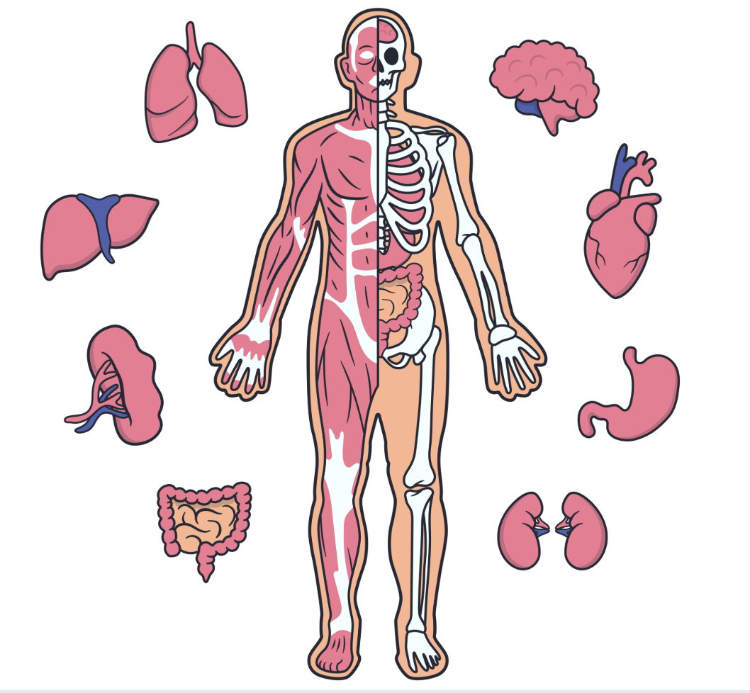
First, we compared a healthy and unhealthy kidney, then a healthy liver with one that was enlarged. Seeing them side by side made the difference obvious in a way no textbook picture could. Second, we focused on the brain, the lungs, and the skull. The skull still had marks from a medical procedure. The medical student explained why bleeding between the brain and the skull is dangerous: the skull is hard bone and cannot give, so the blood has nowhere to go except inward, pressing against the soft brain and causing serious problems. She also walked us through the different regions of the brain and what each one controls. Then she showed us two pairs of lungs, one healthy and one from a smoker. The smoker’s lungs were almost completely black, stained by carbon particles. This station was interesting, but it smelled awful due to the liquid used to preserve the organs.Another medical student lined up a row of hearts, each one having been through a different procedure. For each heart, he explained what had gone wrong and what doctors had done to fix it.
The last lab was my favorite, partly because of who ran it: Olivia Seline’20, now a second-year student. Her station had a full cadaver, a woman who was 79 when she died. Olivia showed us the inside of the knee joint and explained what happens during an ACL tear, then what knee and hip replacements look like, made from a metal and durable medical-grade plastic.
The organs we saw on Tuesday will become chapters in our textbook, and when we get to them, I’ll already know what they really look like. Hopefully, by then, we’ll also be able to name the next picture on the wall on the first try.